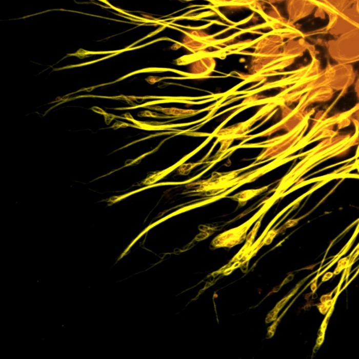
Photomicrograph of the molecular scaffolding of axons.

Photomicrograph of different components of the rat cerebellum, including Purkinje neurons in green, glia (non-neuronal cells) in red, and cell nuclei in blue.

Diffusion MRI image of a patient who has suffered a stroke in the thalamus.
http://www.i-dus.com/2010/11/sel-otak-jika-dilihat-menggunkan.html